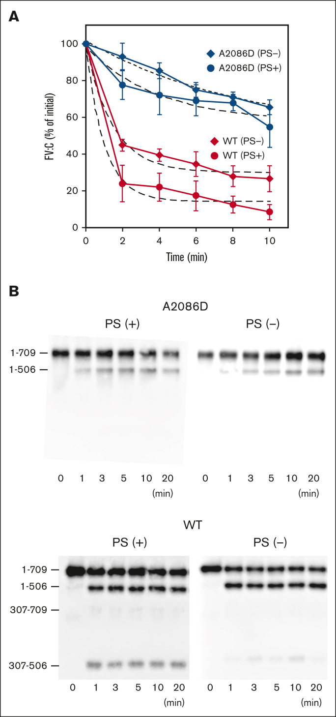
Figure 1.
APC-mediated inactivation and cleavage of FVa-A2086D. (A) APC-mediated inactivation. FV-WT and FV-A2086D (10 nM) were incubated with thrombin (10 nM) for 5 minutes, followed by the addition of hirudin (5 U/mL). FVa (2 nM) was reacted with APC (1 nM) and PL (20 μM) in the presence or absence of PS (30 nM) for the indicated times. After dilution, FVa activity (FVa:C) was measured via a PT-based clotting assay. An initial FV:C was regarded as 100%. Experiments were performed at least 3 times, and average values ± standard deviations are shown. The plotted data were fitted using an equation of single exponential decay (dashed lines). The rate constants (min−1) obtained were FV-WT; 1.02 ± 0.26 (plus PS) and 0.68 ± 0.15 (minus PS), and FV-A2086D; 0.26 ± 0.15 (plus PS) and 0.11 ± 0.05 (minus PS). (B) APC-catalyzed proteolytic cleavage of the HCh of FVa-A2086D. FV-WT and FV-A2086D (5 nM) were incubated with thrombin (5 nM) for 5 minutes, followed by the addition of hirudin (2.5 U/mL). Generated FVa was incubated with APC (1 nM) and PL (20 μM) in the presence or absence of PS (30 nM) for the indicated times. Samples were analyzed on 8% gels, followed by western blotting using an anti-FV HCh mAb 5146 immunoglobulin G.

