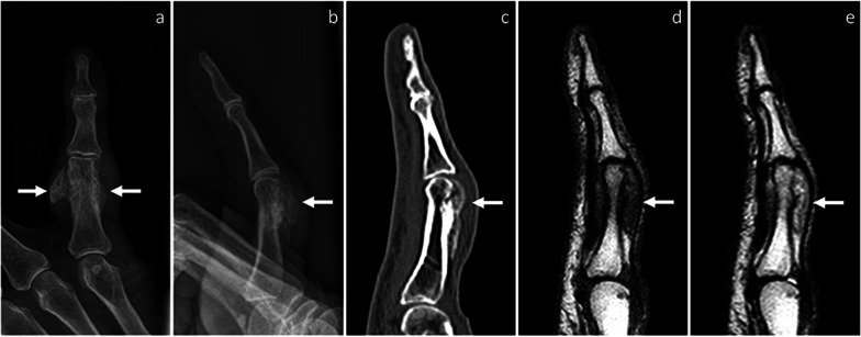
Fig. 1.
BPOP arising from the proximal phalanx of the little finger. On X-rays, frontal (a) and lateral, (b) views show a well-defined mass of heterotopic mineralization, which is contiguous to the proximal phalanx. On sagittal CT image (c), the mass is cortex-based with no cortico-medullary continuity, cortical breakthrough, or marrow extension. On MRI, the mass is hypointense on T1-weighted (d) and hyperintense on T2-weighted (e) sagittal sequences, respectively. Location and imaging findings are in keeping with BPOP. Surgical resection was performed and BPOP was pathologically proven. Arrows point at BPOP in all images

