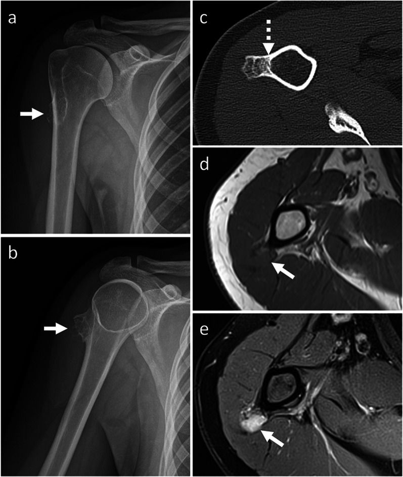
Fig. 9.
Periosteal chondrosarcoma. On X-rays (a, b), a mineralized surface-based mass of the proximal humerus is seen. On axial CT image (c), the mass is cortex-based and partially ossified. Cortical remodeling and erosion (dashed arrow) are noted. On axial T1-weighted (d) and proton density-weighted (e) MRI sequences, the mass shows low and high signal, respectively. No marrow or soft-tissue extension is noted. Periosteal chondrosarcoma was pathologically proven. White arrow points at the lesion in all images

