Fig. 3.
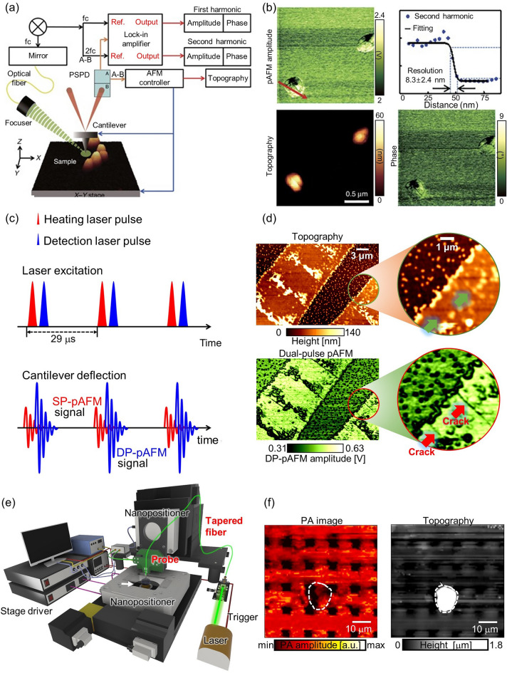
Super-resolution PAM techniques for surface imaging. a Schematic of a super-resolution visible pAFM. b A pAFM image, and topographic and phase images of single gold nanoparticles, with their lateral resolution. c Illustration of the dual-pulse mechanism for DP-pAFM signal generation. d Organic semiconductor topography and a DP-pAFM image for nanocrack analysis. e System configuration of NSPM. f Two-dimensional NSPM image and the topography of a gold microlattice. PAM, photoacoustic microscope; pAFM, photoactivated atomic force microscopy; SP-pAFM, single-pulse photoactivated atomic force microscopy. DP-pAFM, dual-pulse photoactivated atomic force microscopy; NSPM, near-field scanning photoacoustic microscopy; PA, photoacoustic. The images are reproduced with permission from [24, 27, 28]

