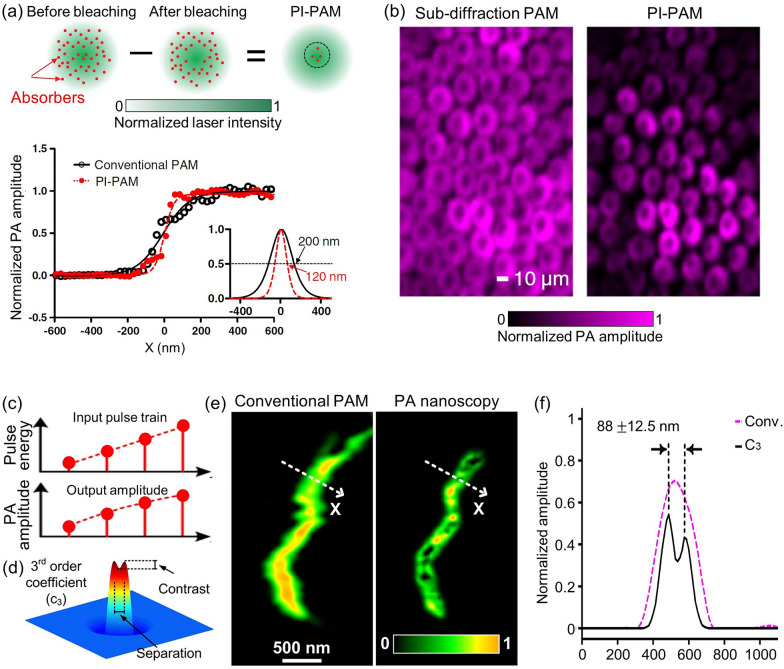
Fig. 4.
Far-field super-resolution PAM techniques for imaging single cells and organelles. a Principle of nonlinear photobleaching-based PI-PAM, depicting the lateral resolutions of PI-PAM and conventional OR-PAM along the edge spread function. b In vitro PI-PAM image of live rose petal epidermal cells. c Illustration of nonlinearity between pulse energy and resultant PA amplitude featured in nonlinear absorption-based PA nanoscopy. d Simulation of a 3rd order PA coefficient image which distinguishes two particles. e A mitochondrion imaged by conventional PAM and 3rd order PA nanoscopy. f Normalized PA amplitude along the dashed lines in panel e. The images are reproduced with permission [25, 26]

