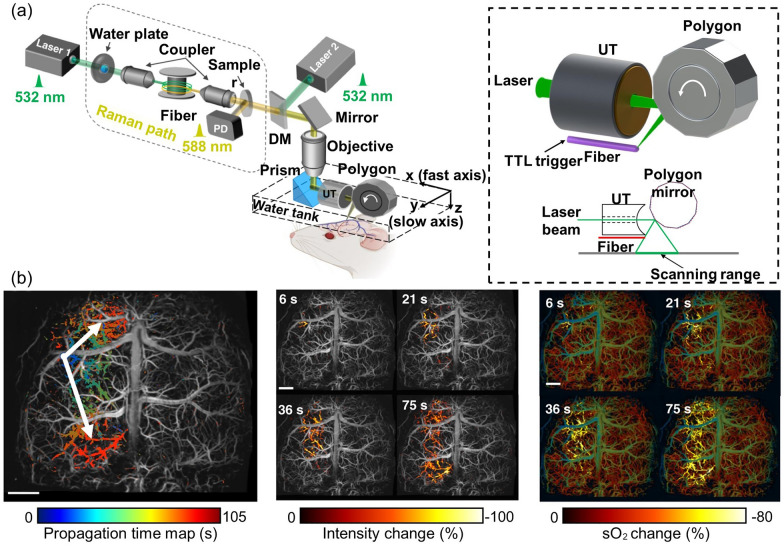
Fig. 6.
Ultrafast wide-field, high-resolution, functional OR-PAM for the study of ischemic stroke. a Schematic of the ultrafast functional PAM. The 532 nm light source and 558 nm Raman source are combined for functional imaging. Inset illustrates the UT, polygon scanner, laser, and scanning range. b Propagation time map of the SD wave and changes in PA intensity and SO2 during the SD wave propagation at different time steps. PAM, photoacoustic microscopy; DM, dichroic mirror; UT, ultrasound; SO2, oxygen saturation; PA, photoacoustic; SD, spreading depolarization. The images are reproduced with permission from [83]

