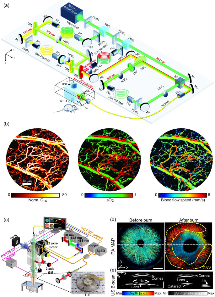
Fig. 7.
High-resolution and functional OR-PAM for the study of cancer. a Schematic of a functional OR-PAM using a five-wavelength pulsed laser and stimulated Raman scattering, b hemoglobin concentration, oxygen saturation, and blood flow speed imaging results from the ear of a tumor-implanted mouse. c–e Quadruple imaging system based on a TUT. c System configuration. d PA depth encoded MAP images and e cross-sectional US B-scan images before and after the chemical burns. OR-PAM, optical-resolution photoacoustic microscopy; CHb, total hemoglobin; SO2, oxygen saturation; HWP, half-wave plate; LPF, long-pass filter; PBS, polarizing beam splitter; M, mirror; FC, fiber coupler; PM-SMF, polarization-maintaining single-mode fiber; SMF, simgle-mode fiber; SPF, short-pass filter; DM, dichroic mirror; NDF, neutral density filter; OL, optical lens; AL, acoustic lens; WT, water tank; UST, ultrasound transducer; BS, beam splitter. PA, photoacoustic; US, ultrasound; TUT, transparent ultrasound transducer; CMOS, complementary metal oxide semiconductor; SLED, superluminescent light-emitting diode; SPECT, spectrometer. The images are reproduced with permission from [17, 142]

