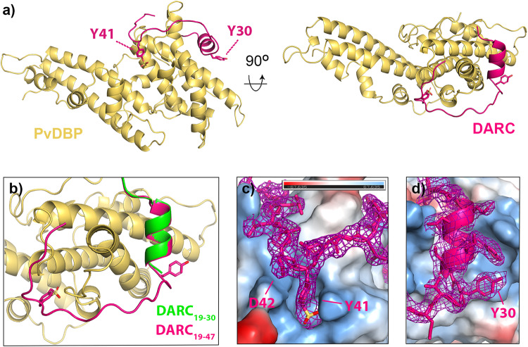
Fig. 1. The structure of PvDBP-RII bound to sulfated DARC ectodomain.
a The structure of PvDBP-RII (yellow) bound to the DARC ectodomain (pink). Residues Y30 and Y41 from DARC are highlighted as sticks, with sulfur in yellow and oxygen in red. b A close up of the DARC ectodomain, showing residues 19-47 of the sulfated ectodomain coloured as a), overlayed with residues 19-30 of the non-sulfated ectodomain in green (from PDB: 4NUV)25. c A close up of residue 41 of DARC, with DARC and the electron density surrounding DARC in pink and PvDBP-RII as a surface coloured by electrostatics (blue as positive charge and red as negative, estimated in pymol). d A close up of residue 30 of DARC, with DARC and the electron density surrounding DARC in pink and PvDBP-RII as a surface coloured by electrostatics (blue as positive charge and red as negative, estimated in pymol). In both c, d, the region of the 2FO-FC map within 2 Å of DARC is shown at a contour level of 1.1.

