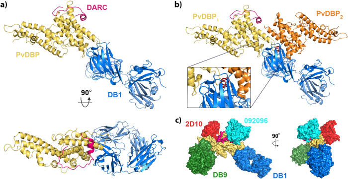
Fig. 4. Structural basis for neutralising antibody binding to PvDBP.
a Structure of PvDBP-RII (yellow) bound to DARC (pink) and antibody DB1. b A model of the putative PvDBP dimer (yellow and orange) bound to DARC and DB1, derived from PDB: 4NUV, showing that DB1 clashes with the putative dimerisation interface. c A composite structure in which four different neutralising antibodies, DB1 (blue), DB9 (green)13, 2D10 (red)32 and 092096 (cyan)20 are docked on to the structure of PvDBP-RII (yellow) and DARC (pink).

