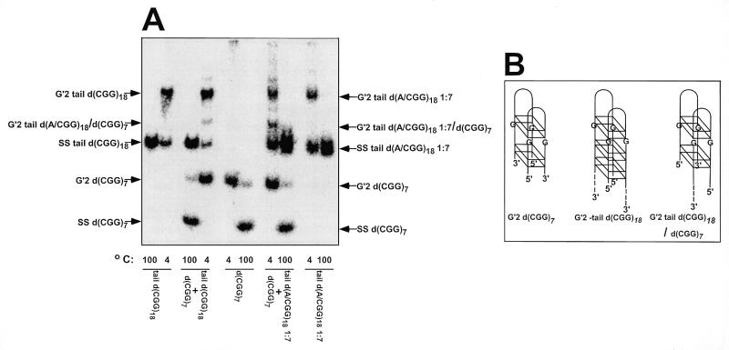
Figure 1.
Stoichiometry of tetraplex structures of tail d(CGG)18 and tail d(A/CGG)18 1:7. Tetrahelical DNA structures were formed at 55°C for 20 h in mixtures that contained in a final volume of 10 µl of TE buffer, 500 mM NaCl and 55 µM of 32P-5′-labeled d(CGG)7, tail d(CGG)18 or tail d(A/CGG)18 or mixtures of 25 µM d(CGG)7 with 25 µM tail d(CGG)18 or tail d(A/CGG)18 1:7. The reaction mixtures were diluted 10-fold in 25 mM Tris–HCl buffer pH 8.0, 1 mM EDTA, 0.5 mM DTT, 20% glycerol and electrophoretically-retarded DNA complexes were resolved from rapidly migrating single-stranded oligomers by electrophoresis at 4°C through a non-denaturing 10% polyacrylamide gel containing 50 mM NaCl in 0.5× TBE buffer. Control DNA samples were denatured at 100°C for 10 min prior to electrophoresis to view the migration of single-stranded oligomers. (A) Electrophoretic separation of tetraplex and single-stranded DNA. Arrows mark the positions of bands of single-stranded and tetraplex DNA complexes. Under the reaction conditions employed, 57 and 28% of tail d(CGG)18 or tail d(A/CGG)18, respectively, were accumulated in tetraplex form. Note hybrid complex bands in samples containing 1:1 mixtures of 32P-5′-labeled d(CGG)7 with tail d(CGG)18 or tail d(A/CGG)18 1:7. (B) Scheme of tetraplex structures of d(CGG)7, tail d(CGG)18 and their hybrid. Based on the findings in (A) and on previously described results (36,37), the bimolecular tetrahelices are depicted as dimers of two hairpins bonded by guanine quartets. Shown are G′2 bimolecular tetraplexes without or with a non-d(CGG) single-stranded tail at their 3′ end (dashed lines). Two pairs of stacked guanine quartets are schematically drawn for G′2 d(CGG)7 tetraplex and three in G′2 tail d(CGG)18 to mark the difference in the number of d(CGG) repeats in the two oligomers. The two hairpins are aligned against each other in one of several possible orientations.

