
FIGURE 2.
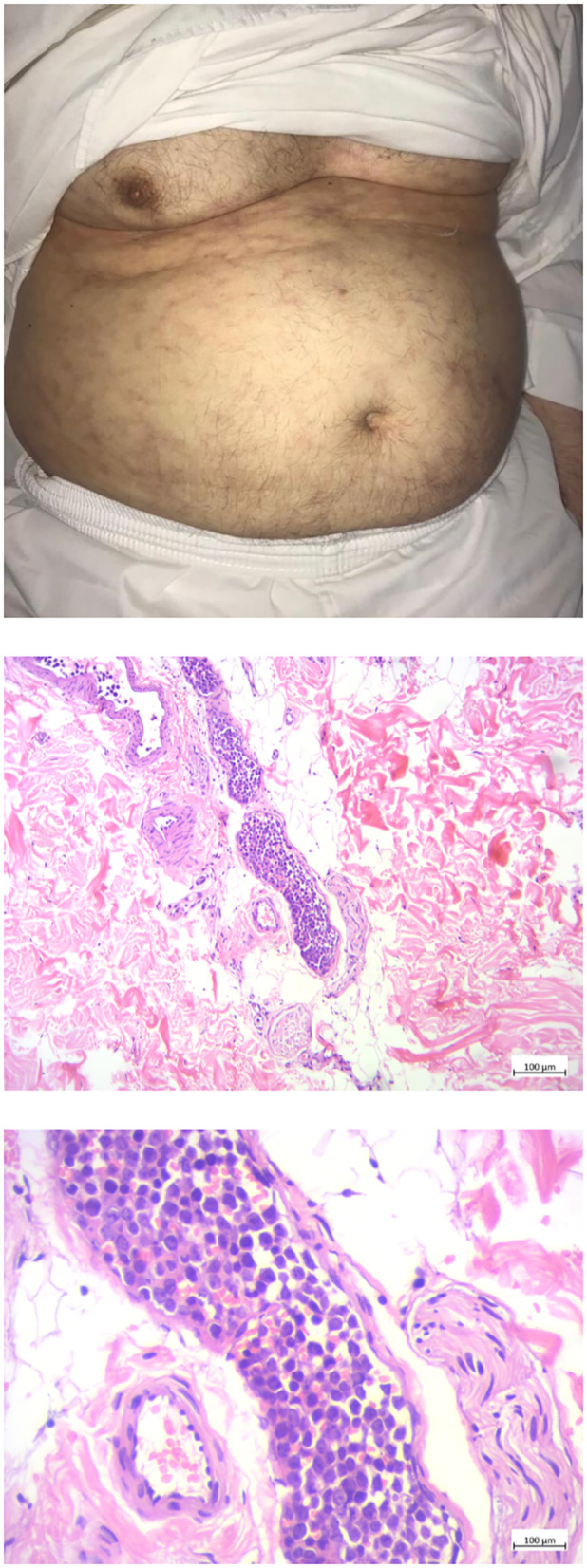
The abdominal skin lesion and the biopsy specimens of abdominal skin lesions; Left: Patient's abdomen upon examination on follow‐up 2 years post‐presentation. The skin shows scattered violaceous telangiectasiae and retiform purpurae. Middle and right: sections show intravascular proliferation of atypical lymphocytic cells inside the dermis. Left: low magnification, right: high magnification.