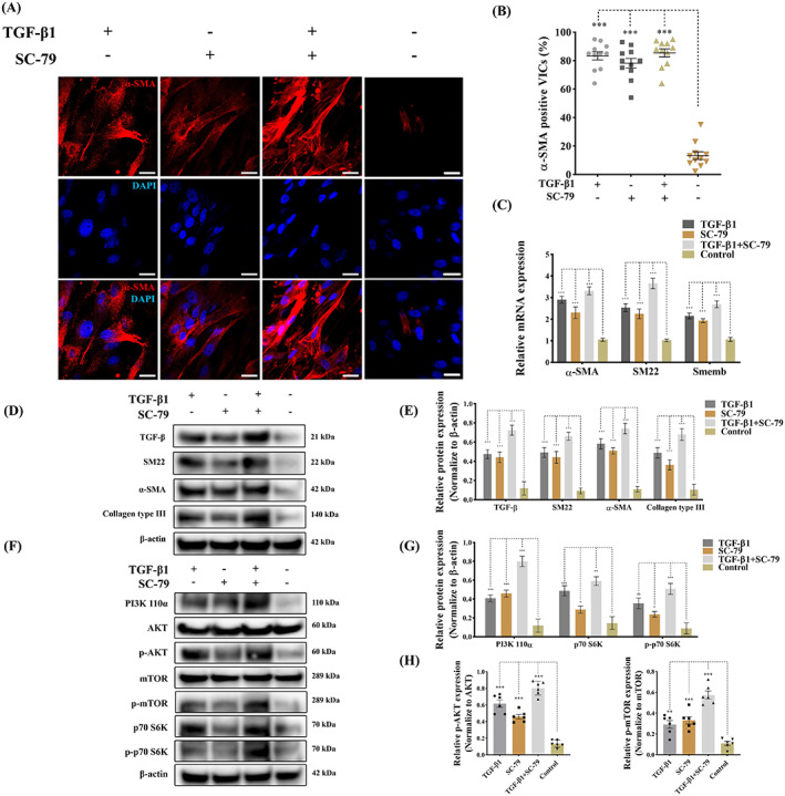
FIGURE 3.

TGF‐β‐induced PI3K activation controls VIC phenotype and ECM protein production. Canine qVICs were exposed to DMSO (Control), TGF‐β1 (10 ng/mL) and SC‐79 (300 nM) treatment. (A, B) Representative confocal images of α‐SMA immunostaining and quantitative analysis of the percentage of α‐SMA positive cells treated with DMSO, TGF‐β1 (10 ng/mL) and SC‐79 (300 nM), scale bar 20 μm (n = 12 microscopic fields). (C) Quantitative RT‐PCR for α‐SMA, SM22 and Smemb mRNA expression in qVICs treated with DMSO, TGF‐β1 (10 ng/mL) and SC‐79 (300 nM) (n = 6). (D, E) Representative western blot of α‐SMA, SM22, collagen type III and TGF‐β and quantitative analysis of the relative protein expression (n = 6). (F, G, H) Representative western blot of PI3K p110α, AKT, phosphorylated AKT (p‐AKT), mTOR, phosphorylated mTOR (p‐mTOR), p70 S6K, phosphorylated p70 S6K (p‐p70 S6K) protein expression and quantitative analysis of the relative protein expression (n = 6). Results are presented as mean ± SEM. ANOVA followed by Tukey's range test. *p < 0.05, **p < 0.01, ***p < 0.001 compared to control. ANOVA, analysis of variance; ECM, extracellular matrix; TGF‐β, transforming growth factor β; VIC, valve interstitial cell.
