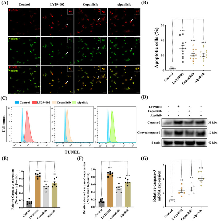
FIGURE 6.

Antagonism of PI3K pathway promotes apoptosis in canine aVICs. aVICs were treated with DMSO (Control), LY294002 (60 μM), copanlisib (5 μM) and alpelisib (50 μM) for 3 days. (A, B) Representative confocal images of TUNEL (red) staining and quantitative analysis of the percentage of TUNEL positive (apoptotic) cells (arrowhead), scale bar 20 μm (n = 12 microscopic fields/treatment). (C) Flow cytometry analysis of TUNEL staining and quantification of the percentage of apoptotic cells at Day 3 (n = 3). (D–F) Representative western blot of caspase‐3, cleaved caspase‐3 and β‐Actin protein expression and quantification of the relative protein expression (n = 6). (G) Quantitative RT‐PCR for caspase‐3 mRNA expression in aVICs treated with PI3K inhibitors after 3 days (n = 6). Results are presented as mean ± SEM. ANOVA followed by Tukey's range test. *p < 0.05, **p < 0.01, ***p < 0.001 compared to control. ANOVA, analysis of variance; aVIC, activated myofibroblast phenotype.
