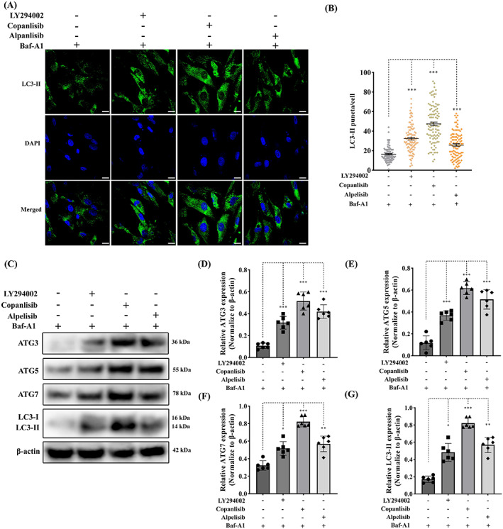
FIGURE 7.

Inhibition of PI3K signalling enhances autophagy in canine aVICs. aVICs were exposed to DMSO (Control), 60 μM LY294002, 5 μM copanlisib and 50 μM alpelisib treatment with 5 μM baflomycin‐A1 (Baf‐A1) for 16 h. (A, B) Representative confocal images of LC3‐II marked autophagosomes (green) and quantitative analysis of the number of LC3‐II puncta, scale bar 20 μm (n = 98 cells/treatment). (C) Representative western blot of ATG3, ATG5, ATG7 and LC3‐II protein expression in aVICs exposed to DMSO and PI3K inhibitors. (D–G) Quantification of the relative protein expression of ATG3, ATG5, ATG7 and LC3‐II (n = 6). Results are presented as mean ± SEM. ANOVA followed by Tukey's range test. *p < 0.05, **p < 0.01, ***p < 0.001 compared to control. ANOVA, analysis of variance; aVIC, activated myofibroblast phenotype.
