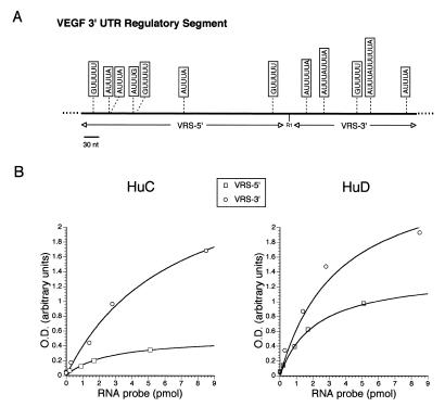
Figure 3.
RNA-binding analysis of HuD and HuC with truncated portions of the VRS. (A) Schematic diagram of the VRS highlighting the motifs which have been linked to ELP–RNA binding (3–5). The truncated VRS probes are shown below the diagram. (B) RNA-binding curves for HuC and HuD to the truncated VRS probes.

