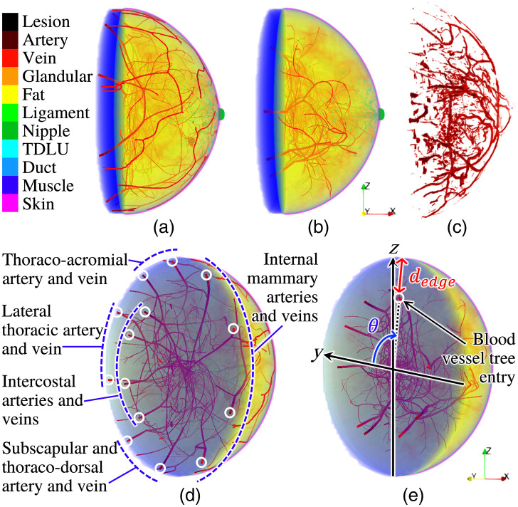
Fig. 2.
Blood vessels in an NBP (type B, left breast) with (a and d) and without (b and e) blood vasculature customization and (c) a clinical OAT image acquired by TomoWave Laboratories employing LOUISA-3D3 at the MD Anderson Cancer Center and postprocessed to extract blood vascular structures.33 Paraview40 was used for volume rendering.

