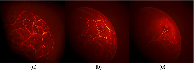
Fig. 8.
Visual comparison of 3D OAT breast images reconstructed from (a) clinical measurement data and simulated measurement data produced using (b) the proposed NBP and (c) the unmodified VICTRE NBP. In panel (a), the clinical data were acquired by TomoWave Laboratories using LOUISA-3D.3 In the unmodified VICTRE NBP, tissue properties were assigned to each tissue type in a piecewise constant manner. All three images were reconstructed using the FBP method.42 The images were visualized using Paraview’s volume rendering technique, which accumulates intensities based on the selected color and opacity maps.40

