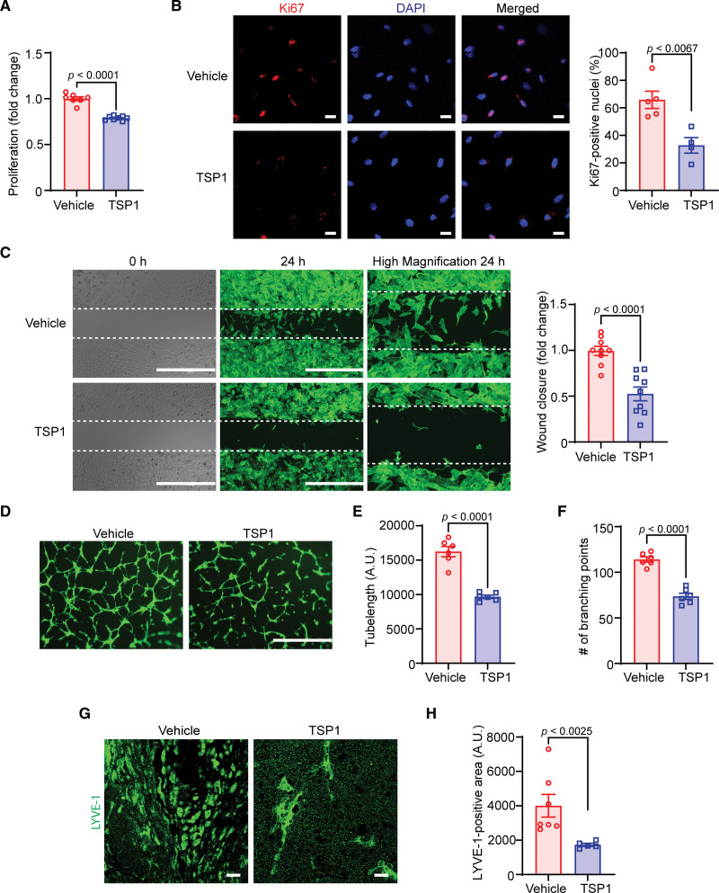
Figure 2.
TSP1 (thrombospondin-1) suppresses both in vitro and in vivo lymphangiogenesis. A, Vehicle- and TSP1-pretreated (22 nM, 4 h) human lymphatic endothelial cells (LECs) were stimulated with VEGF (vascular endothelial growth factor)-C (100 ng/mL) and proliferation investigated after 48 hours using the WST (water-soluble tetrazolium)-1 assay. Data are representative of 3 independent experiments performed at least in duplicate. B, LECs plated on coverslips were pretreated and stimulated as in A for 24 hours. Cells were immunostained for Ki67 (red) and nuclei counterstained with DAPI (4’,6-diamidino-2-phenylindole; blue). Images of at least 4 random microscopic fields were captured. Representative images are shown. Scale bar, 20 µm. Bar graph represents the percentage of Ki67-positive nuclei (n=4–5). C, LEC migration in response to vehicle (VEGF-C) and TSP1 (VEGF-C+TSP1) was investigated after 24 hours using Culture-Insert 2 Well 24 (ibidi USA). Representative images of wounds at 0 and 24 hours are shown. Scale bar, 1000 μm. Bar diagram shows quantification of wound closure (n=9). D, Vehicle- or TSP1-pretreated LECs were seeded in wells of a Matrigel-coated plate in basal medium containing VEGF-C±TSP1 and tube formation determined after 6 hours. Representative images of tube formation are shown. Scale bar, 1000 µm. Images of random fields were captured and tube length (E) and number of branching points (F) quantified (n=6). G and H, Wild-type male mice were injected SC with Matrigel solutions premixed with either VEGF-C or VEGF-C+TSP1. Plugs were isolated after 10 days, sectioned, and immunostained for LYVE-1 (lymphatic vessel endothelial hyaluronan receptor-1). G, Representative images of LYVE-1 staining of cross-sections of the Matrigel plugs are shown. Scale bar, 20 μm. H, Quantification of LYVE-1–positive area (n=5–7). Statistical analyses were performed using a 2-tailed unpaired Student t test (A–C, E, and F) and a Mann-Whitney U test (H). Data represent mean±SEM.

