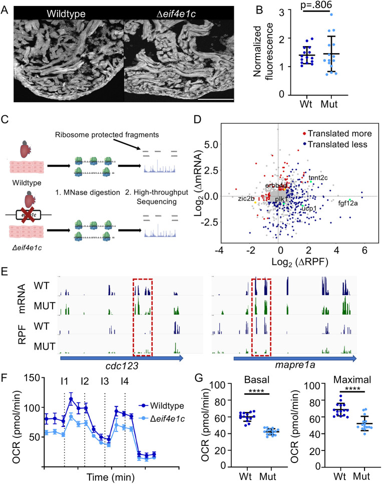
Fig. 4.
Ribosome profiling indicates translational changes in eif4e1c mutant hearts. (A) Example of fluorescence in the hearts of OPP-injected fish. Scale bar: 100 µm. (B) Quantification of OPP fluorescence shows no significant difference between mutant and wild-type fish (normalized fluorescence average wild type=140,279 arbitrary density units (adu)/µm2, mutant=145,096 adu/µm2; Mann–Whitney, P=0.806; n=15). Error bars represent mean±s.e.m. (C) Diagram of the ribosome profiling method. Ribosome-bound mRNA is purified from wild-type and mutant hearts and then subjected to micrococcal nuclease (MNase) digestion. Fragments of mRNA protected by being bound by ribosomes are then purified and subjected to high-throughput sequencing. (D) Differences (mutant/wild type) in mRNA abundance (log2) are plotted versus changes in the abundance of ribosome-protected fragments (RPF). Genes for which translation does not change significantly are plotted in gray, genes for which translation is decreasing in the mutant are plotted in red and genes for which translation is increasing in the mutant are plotted in blue. Decreasing genes mentioned in the text are colored gold and increasing genes mentioned in the text are colored green. (E) High-throughput sequencing browser tracks for two genes (blue arrows) that are involved in cell-cycle progression (cdc123 and mapre1a). Wild-type (WT) data is shown in blue, and mutant (MUT) data is shown in green. The top two tracks (mRNA) show the abundance of mRNA (as measured from RNAseq) and the bottom two tracks show the abundance of RPFs. Dashed red boxes highlight decreasing levels of RPF where mRNA levels are comparable. (F) Time course of oxygen consumption rates (OCR) from mitochondria measured by the Seahorse analyzer. Dotted lines indicate time points of drug injection. Readings were normalized to the number of mitochondria using qPCR. The first injection (I1) is of ADP to stimulate respiration, the second injection (I2) is of oligomycin to inhibit ATP synthase (complex V) decreasing electron flow through transport chain, the third injection (I3) is of FCCP to uncouple the proton gradient, and the fourth injection (I4) is of antimycin A to inhibit complex III to shut down mitochondrial respiration. Error bars represent mean±s.e.m. (G) Basal respiration is calculated as the OCR average after addition of ADP (I1 to I2) subtracting the non-mitochondrial respiration after injection of antimycin A (after I4) (mean wild type=60.01, mutant=42.25; Welch's t-test, ****P<0.0001; n=15). Maximal respiration is calculated as the OCR average after FCCP addition (I3 to I4) subtracting the non-mitochondrial respiration after injection of antimycin A (after I4) (mean wild type=68.73, mutant=52.19; Welch's t-test, ****P<0.0001; n=15). Error bars represent mean±s.e.m. Mut, mutant; Wt, wild type.

