FIGURE 4.
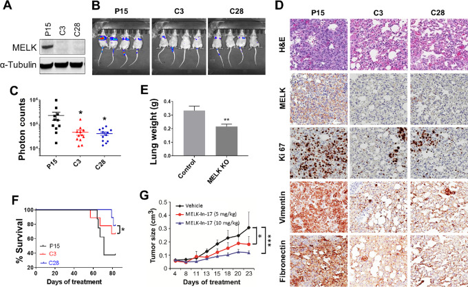
Effect of inhibition of MELK signaling on tumor growth and lung metastasis in TNBC xenograft mouse models. A, MELK expression in Cas9-p15 (P15) and MELK KO MDA-MB-231 (C3 and C28) cells. α-Tubulin was used as a loading control. B–F, MELK KO significantly suppressed lung metastases in an MDA-MB-231 xenograft mouse model. Female athymic nude mice were injected intravenously with luciferase-transfected Cas9-p15 or MELK KO MDA-MB-231-Luc-GFP stable cells. Metastatic tumors were measured weekly for 7 weeks by whole-body luciferase imaging using an IVIS 100 Imaging System. Shown are mouse whole-body luciferase images (B), photon counts per area (C), lung weight per mouse measured at week 7 following cell inoculation (D), mouse OS over a period of 80 days following cell inoculation (E), and IHC staining for hematoxylin and eosin (H&E), MELK, proliferation (Ki67), and mesenchymal markers (vimentin and fibronectin; F) in mice inoculated with Cas9-p15 or MELK KO MDA-MB-231 cells. Images were taken under 200 × magnification. G, MELK-In-17 significantly suppressed tumor growth in a 4T1 xenograft mouse model (P < 0.05). Murine 4T1 TNBC cells were injected into the mammary fat pads of female BALB/c mice. When tumor size reached 75–150 mm3, grouped mice were treated with vehicle or MELK-In-17 at 5 or 10 mg/kg via intraperitoneal injections daily for 25 days. In C–G, data are presented as mean ± SD. *, P < 0.05; **, P < 0.01.

