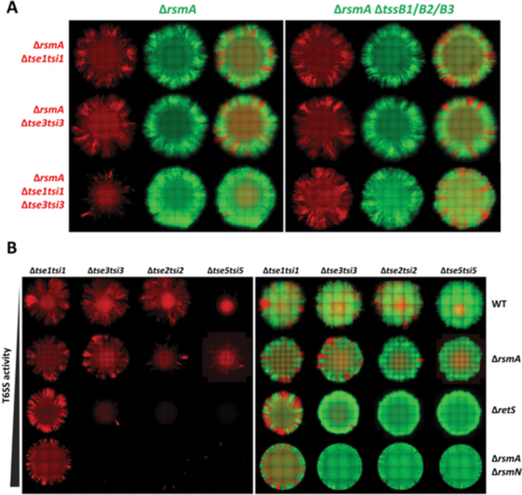
Fig 4. T6SS toxin potency differs and increases with elevation in T6SS activity or synergistic toxin action.
(A) Representative fluorescent images of ΔrsmA strains sensitised to Tse1 or Tse3 individually and in combination. Both single fluorescence channel and overlay images showing distribution of sensitised prey in a mix with T6SS+ and subsequently T6SS- (ΔtssB1ΔtssB2ΔtssB3) parental strain. Strains contain constitutively expressed fluorescent proteins, prey labelled with mCherry (shown in red) and attacker with sfGFP (shown in green). All bacteria mixed at 1:1 ratio, inoculum OD600 = 1.0, grown for 48h at 37°C on LB with 2% (w/v) agar. (B) Whole colony fluorescent microscopy image of T6SS sensitive strain distribution within a colony when mixed with a T6SS+ strain. Corresponding fluorescent channel composite images showing toxin sensitive bacteria in red and T6SS+ parental strain in green. Strains sensitised to individual H1-T6SS toxins in each of the columns in the following order: Tse1, Tse3, Tse2, and Tse5. Each row contains strains with different regulatory background in following order of ascending H1-T6SS activity: WT, ΔrsmA, ΔretS, and ΔrsmAΔrsmN. Strains contain constitutively expressed fluorescent proteins, prey labelled with mCherry (shown in red) and attacker with sfGFP (shown in green). All bacteria mixed at 1:1 ratio, inoculum OD600 = 1.0, grown for 48h at 37°C on LB with 2% (w/v) agar.

