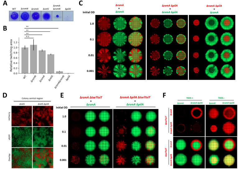
Fig 6. Loss of T4P hinders P. aeruginosa ability to intermix within communities resulting in impaired T6SS killing.
(A, B) Twitching assay assessing surface motility of mutants in the Gac/Rsm pathway and ΔpilA mutant strains as compared to WT. Images comparing strain twitching zones as seen by crystal violet staining (A) and corresponding quantification of the relative twitching zone area (B). (Mean + SD of n = 5 biological repeats). (C) A set of mixed bacterial macrocolonies consisting of T4P+ (ΔrsmA) and T4P- (ΔrsmAΔpilA) strains tagged with constitutive mCherry (red) and sfGFP (green). Single fluorescence channel and overlay images of T4P+ and T4P+, T4P- and T4P-, T4P- and T4P+ mixed colonies, displayed to scale. (D) Bacterial T4P+ (ΔretS) and T4P- (ΔretSΔpilA) mutant intermixing at single-cell scale. Mix of isogenic strains with mCherry and sfGFP tags grown in the interface between media agar and glass coverslip until dense community is established. Single fluorescence channel and overlay images show the different extent of local intermixing. (E) A set of mixed bacterial communities where TseT sensitive prey growth is spatially restricted in presence of T6SS+ (ΔrsmA) parental strain. Showing both individual fluorescent channel and overlay images of both prey population in red and attacker in green. Toxin sensitised strain mixed with a parental strain at 1 to 1 ratio and after adjusting inoculum density (OD600 1.0; 0.1; 0.01; 0.001) spotted on LB agar to assess how reduction in lineage intermixing impacts T6SS mediated competition. (F) TseT-mediated competition was used to assess impact on T6SS mediated killing in a mix of T4P+ and T4P- bacteria. The panel contains individual red channel images showing toxin sensitive prey distribution in presence of T6SS+ (left) or T6SS- (right) attacker strain in the upper two rows. Corresponding overlay images showing distribution of both toxin sensitive prey (in red—mCherry) and T6SS attacker population in green (sfGFP) shown in the lower two rows. T4P+ prey (ΔrsmAΔtseTtsiT) strains in the upper row, with T4P- prey (ΔrsmAΔpilAΔtseTtsiT) in the lower row. With T4P+ attacker in the first column and T4P- attacker strains in the second. All bacteria mixed at 1 to 1 ratio, with inoculum density as specified for (C) and (E), or OD600 1.0 for (F), grown for 48h at 37°C on LB with 2% (w/v) agar for (C) and grown at 25°C on LB with 1.2% (w/v) agar for (E, F). 1 of 1 biological repeat shown for all images containing ΔpilA mutant strains.

