Figure 6.

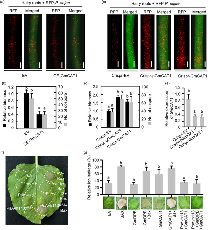
PsAvh113 inhibits the positive immune regulator GmCAT1‐induced cell death. (a–d) Confocal microscopy analysis (a, c) and quantification (d) of oospores and pathogen biomass in soybean hairy roots overexpressing WT GmCAT1 (a, b) or edited GmCAT1 (c, d) along with the EV control. Scale bar, 0.25 mm. Confocal microscopy analysis was conducted at 48 hpi. In (b) and (d), black columns represent biomass, and grey columns represent oospore count. (e) The transcript levels of GmCAT1 in GmCAT1‐edited hairy roots. In (b–e), data represent mean ± SE, and different letters indicate statistically significant differences (P < 0.01; Duncan's multiple range test). (f) Overexpression of PsAvh113 in N. benthamiana suppresses Bax‐triggered cell death. N. benthamiana leaves were infiltrated with A. tumefaciens containing PVX‐PsAvh113, PVX‐PsAvh113▵IR2, PVX, or PVX‐Avr1b, either alone or with A. tumefaciens cells carrying PVX‐Bax, which were infiltrated 24 h later. EV, empty vector. (g) GmCAT1 induces cell death in N. benthamiana leaves, whereas overexpression of PsAvh113 in N. benthamiana suppresses PCD triggered by GmCAT1. Pictures were taken at 5 dpi. Experiments were repeated three times with similar results.
