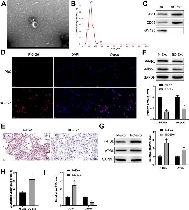
Figure 2.
BC cell-derived exosomes facilitate fat loss in adipocytes. (A) Exosomes isolated from BC cells were observed with a transmission electron microscope. (B) Particle size analysis of the isolated exosomes. (C) Western blotting was used to show the expression of CD81, CD63 and GM130. (D) 3T3-L1 cells was co-incubated with PKH-26-marked BC cells and observed with a fluorescence microscope. (E) Oil Red O staining was used to show lipid droplet accumulation in adipocytes. (F, G) Western blotting was used to quantify the expression of PPARγ and AdipoQ as well as P-HSL and ATGL in adipocytes. (H) The levels of free glycerol released by adipocytes within 24 h. (I) The mRNA levels of UCP1 and leptin were quantified by RT-qPCR. Measuring data were expressed as mean ± standard deviation. Comparisons between groups were performed using the t-test. *P < 0.05.

