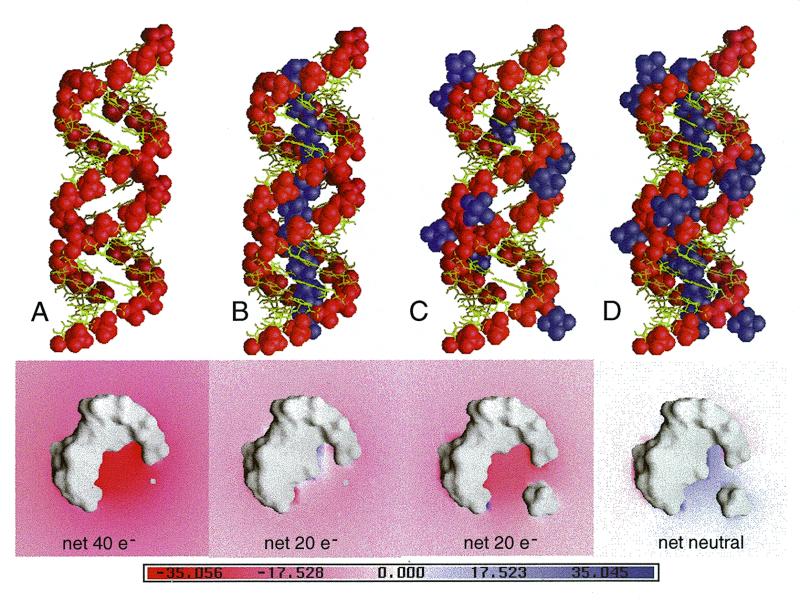
Figure 5.
Electrostatic surface potential of an idealized RNA duplex without [(A), bottom] and with [Mg(H2O)6]2+ ions placed inside the deep groove [(B), bottom], with [Mg(H2O)6]2+ ions at the outer mouth of the groove [(C), bottom] and inside the deep groove plus at the outer mouth of the groove [(D), bottom]. The idealized poly(rG-rU) RNA model was constructed by repeating the A-form conformation of the G3pT4/A17pC18 pairs of the r(GCG)d(TATACGC) crystal structure with the idealized O2′ atoms added. The Mg II site (Table 2) is also repeated in the deep groove of the idealized helix for subsequent electrostatic calculation. The colors indicate the scale for the electrostatic potential ranging from –35 kT/e (red) to +35 kT/e (blue). The locations of the phosphates and the [Mg(H2O)6]2+ ions are indicated in the top panels as red and purple spheres, respectively. (See Materials and Methods for more details.)

