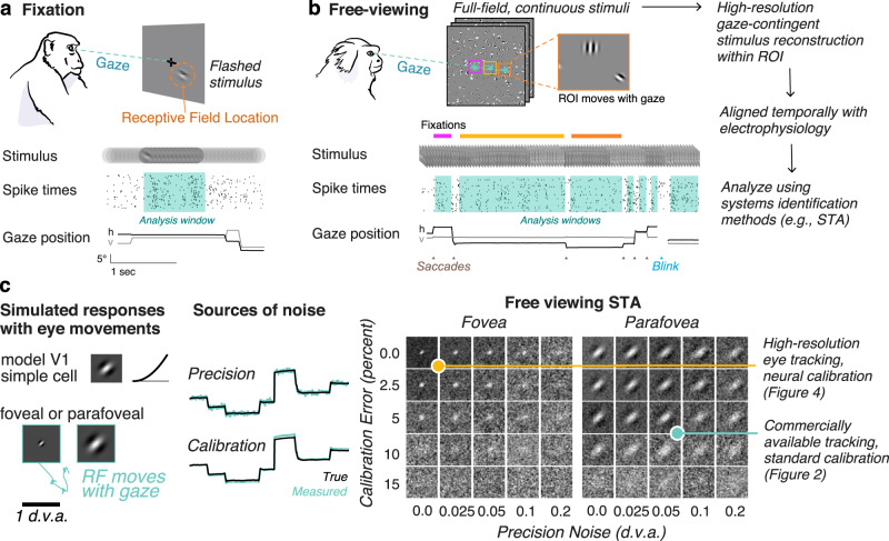
Fig. 1. Free-viewing paradigm and gaze-contingent neurophysiology.
a Conventional fixation paradigm with flashed stimuli. Spike times are aligned to stimulus onset and analyzed during a window during fixation. b Free viewing: subjects freely view continuous full-field stimuli. Shown here is dynamic Gabor noise. All analyses are done offline on a gaze-contingent reconstruction of the stimulus with a region of interest (ROI). Analysis windows are extracted offline during the fixations the animal naturally produces. c Simulation demonstrates how uncertainty in the gaze position (due to accuracy and precision) would limit the ability to map a receptive field. (Left) A model parafoveal simple cell (linear receptive field, half-squaring nonlinearity, and Poisson noise) moves with the gaze. Example gaze trace shown in cyan. RF inset is 1 d.v.a. wide. (Middle) errors in precision are introduced by adding Gaussian noise to the gaze position. Errors in calibration are introduced by a gain factor from the center of the screen. (Right) The recovered RF using spike-triggered averaging (STA) on a gaze-contingent ROI. Of course, with zero precision noise or calibration error, the STA recovers the true RF. Adding either source of noise degrades the ability to recover the RF, however, some features are still recoverable for a wide range of noise parameters at this scale. RFs that are smaller or tuned to higher spatial frequencies require high-resolution eye tracking. The yellow and cyan dots indicate two levels of accuracy that are explored in Figs. 2 and 4, respectively. Animal drawings in panel a and b were created with help from Amelia Wattenberger.

