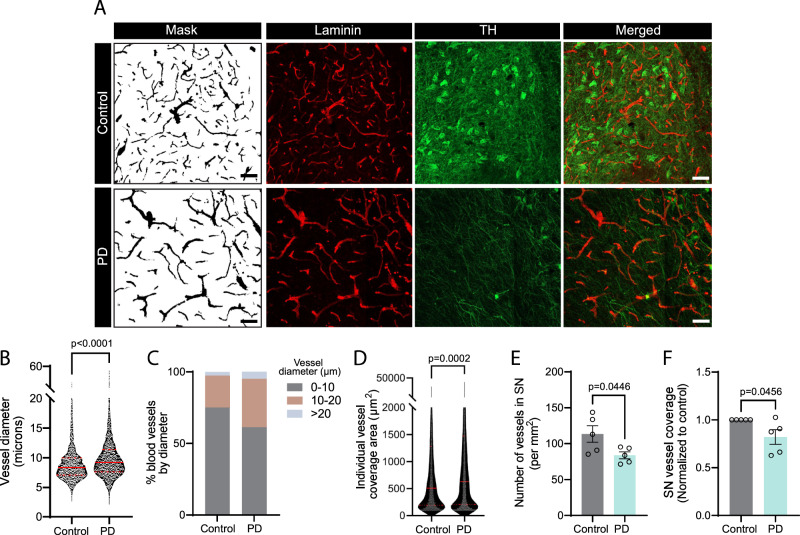
Fig. 8. The BBB chip recapitulates morphological changes to the vasculature observed in the substantia nigra of PD patients.
A Confocal images of human postmortem sections of the substantia nigra immunostained for laminin (red) and TH (green). The brain sections were obtained from age- and sex-matched controls or patients with PD. A mask of the laminin-positive staining was produced using FIJI image analysis software. B–F Graphs reporting laminin-positive vessel diameter (B) and size distribution (C), individual laminin-positive vessel coverage area (D), number of laminin-positive vessels (E), and overall vessel coverage (F) in the substantia nigra of PD patients vs control. Data were collected using postmortem brain sections originating from five age- and sex-matched controls and five PD cases; error bars represent mean + SEM. Violin plot in (B) and (D) indicate the median (red line) and quartile (red dotted line) values. Statistical analysis was performed using two-tailed unpaired Student’s t test with equal s.d. Scale bar: 100 µm (A). Source data are provided as a Source Data file.

