
FIGURE 3.
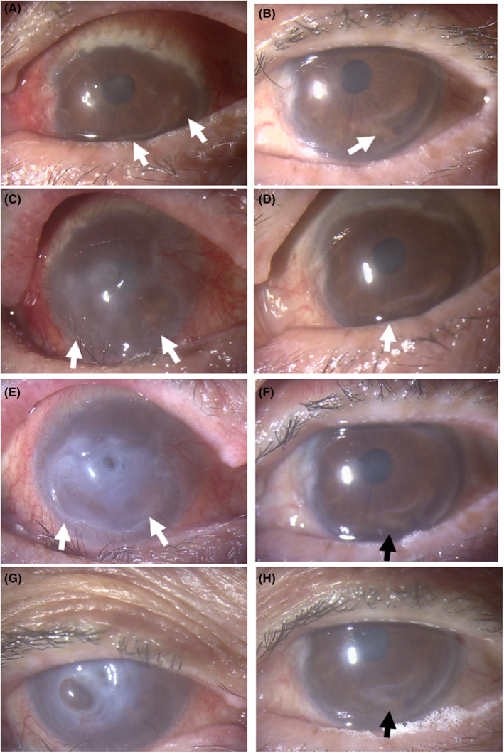
Biomicroscopic images. Diffuse scleritis with massive peripheral corneal infiltration and crescent‐shaped corneal ulcer (arrows) on the nasal to inferior side in the right eye (A) and crescent‐shaped corneal ulcer (arrow) on the temporal to inferior side in the left eye (B) at referral to an ophthalmologist. In a month, note central corneal haze and infiltrates with inferior crescent‐shaped wide ulcer (arrows) in the right eye (C) and peripheral corneal infiltrates with inferior ulcer (arrow) in the left eye (D). In the following 3 weeks, the central cornea has been perforated in the right eye with the inferior wide ulcer (arrows, E) while the corneal ulcer is stable in the left eye (arrow, F). Four months after the referral, the right eye is stable with corneal opacity with iris incarceration (G) and the left eye is stable with healed corneal ulcer (H).