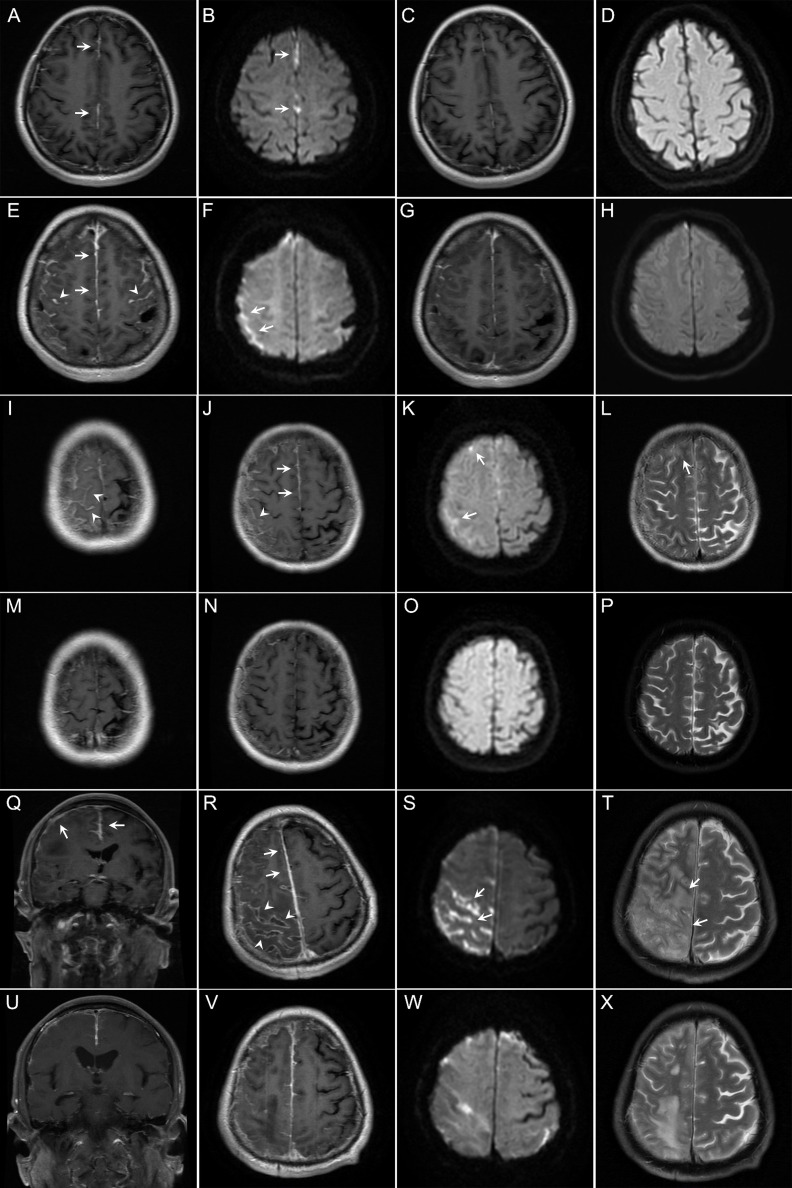
Figure 2.
Findings on magnetic resonance imaging (MRI) of the brain in the patients with rheumatoid meningitis in this study. (A–D) In case 1, brain contrast MRI performed 4 months after onset of symptoms of meningitis showed pachymeningeal enhancement (A, arrows). Diffusion-weighted imaging (DWI) showed restricted diffusion (B, arrows). The patient experienced improvement, with resolution of the lesions 10 months after immunotherapy (C, D). (E–H) In case 2, brain contrast MRI performed 5 months after onset of symptoms of meningitis showed enhancement of both the pachymeninges (E, arrows) and leptomeninges (E, arrowheads). Restricted diffusion was shown on DWI (F, arrows). Repeated MRI showed reduction of contrast enhancement and resolution of restricted diffusion 3 months after immunotherapy (G, H). (I–P) In case 4, brain MRI performed 12 months after onset of symptoms of meningitis also showed involvement of both the pachymeninges (J, arrows) and leptomeninges (I, J, arrowheads) with restricted diffusion (K, arrows). T2-weighted MRI scans showed a small cortical lesion (L, arrow). Repeated MRI showed resolution of the lesions 28 months after immunotherapy (M–P). (Q–X) In case 5, brain contrast MRI performed 3 months after onset of symptoms of meningitis revealed asymmetric involvement of both the pachymeninges (Q, R, arrows) and leptomeninges (R, arrowheads). Note the lesions are mainly located on the convex surface of the cerebral hemisphere. DWI showed sulcal restricted diffusion (S, arrows). T2-weighted MRI scans showed lesions in the parenchyma (T, arrows). Repeated MRI showed reduction of the lesions 3 months after immunotherapy (U–X).

