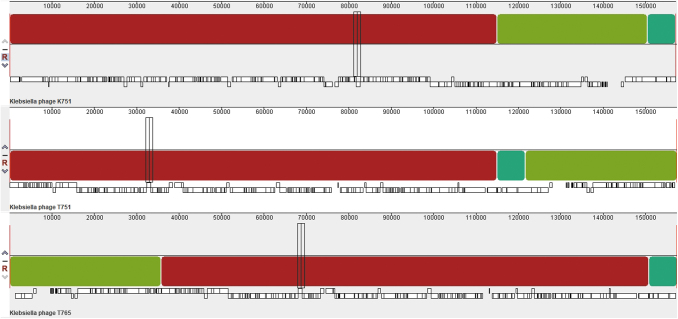
FIG. 2.
Progressive multiple alignment performed with MAUVE to compare Klebsiella_virus_K751, Klebsiella_virus_T751, and Klebsiella_virus_T765. Each genome is arranged horizontally with homologous segments (locally collinear blocks) delineated as colored rectangles. Regions inverted with respect to the first genome taken as reference are placed below those that match in forward orientation.

