Figure 1.
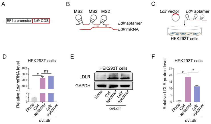
Construction of the Ldlr expression vector and aptamer: (A) Schematic illustration of Ldlr-expressing vector construction. The CDS of Ldlr was cloned downstream of the EF1α promoter. (B) Schematic illustration of the interaction between Ldlr mRNA and Ldlr aptamer. The Ldlr aptamer contains 3 MS2 stem-loop sites and a sequence region base-pair matched with Ldlr mRNA near the translation initiation site. (C) Schematic illustration of co-transfection of Ldlr-expressing vector (ovLdlr) and Ldlr aptamer into HEK293T cells (D) Expression of Ldlr mRNA in HEK293T cells treated as indicated. Gapdh served as the internal control. (E) Western blot analysis of LDLR protein in HEK293T cells with indicated treatments. GAPDH expression served as the loading control. The data shown are representative of 3 independent experiments. (F) Quantification of Western blot band intensity by densitometry. Data are presented as mean±SEM of 3 independent experiments. *P<0,05 by one-way ANOVA. ns, no significance.

