Fig. 5.
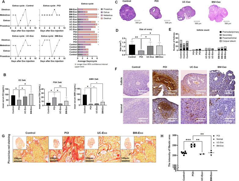
Ovarian function restoration in the POI mouse model after exosome treatment. The therapeutic effect in the POI mouse model was compared between the healthy control (control), untreated POI (POI), umbilical-derived MSC exosome treatment (UC-Exo), and bone marrow-derived MSC-derived exosome treatment (BM-Exo) groups. A Estrous cycle after exosome treatment by daily vaginal smear analysis. Animal shows significantly delayed cycle (> 95% CI) is highlighted with ǂ symbol. B Average serum E2, AMH, and FSH levels in the control, POI, UC-Exo, and BM-Exo groups at 2 weeks after treatment. C Representative image of ovarian tissue with H&E staining. D Average size of the ovaries in the control, POI, UC-Exo, and BM-Exo groups at 2 weeks after treatment. E Number of ovarian follicles in the control, POI, UC-Exo, and BM-Exo groups at 2 weeks after treatment. F TUNEL assay in ovarian tissue among the control, POI, UC-Exo, and BM-Exo groups at 2 weeks after treatment. G, H Picrosirius Red staining assay in ovarian tissue among the control, POI, UC-Exo, and BM-Exo groups at 2 weeks after treatment (small image magnification: 50×; large image magnification: 400×). Data are presented as the mean ± SD. (n = 3, significance level, *p < 0.05, **p < 0.005, ***p < 0.0005, ****p < 0.0001; NS: Not significant.)

