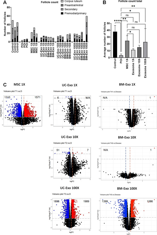
Fig. 7.

Differences between MSC treatment and exosome treatment in the POI mouse model. A Number of ovarian follicles in the healthy control (control), untreated POI group (POI), MSC treatment group (MSC 1×), equal amount of exosomes (UC-Exo 1×, BM-Exo 1×), tenfold higher amount of exosomes treatment (UC-Exo 10×, BM-Exo 10×), and 100-fold higher amount of exosomes treatment (UC-Exo 100×, BM-Exo 100X). B Comparison of the average number of total follicles among the control, POI, MSC 1×, average UC-Exo and BM-Exo (exosomes 1×, 10×, and 100×) groups. C Altered gene expression by RNA-seq analysis in each treatment compared with untreated POI mouse ovaries. A volcano plot shows significantly increased genes (blue) and significantly decreased genes (red) after MSC and exosome treatment. Data are presented as the mean ± SD. (n = 3, significance level, *p < 0.05, **p < 0.005, ***p < 0.0005, ****p < 0.0001; NS: Not significant.)
