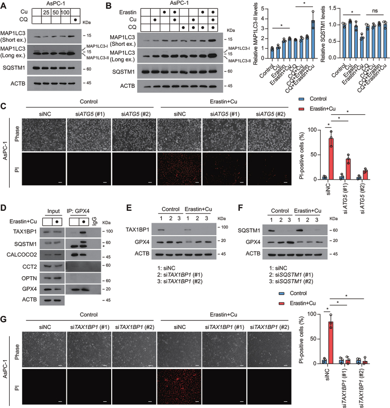
Figure 3.

TAX1BP1 is essential for GPX4 degradation during ferroptosis. (A) Western blot analysis of the expression of indicated proteins in AsPC-1 cells after treatment with dose-dependent copper sulfate (Cu) for 12 h in the absence or presence of chloroquine (CQ, 20 μM). (B) Western blot analysis of the expression of indicated proteins in AsPC-1 cells after treatment with copper sulfate (Cu, 50 μM) and erastin (1.25 μM) for 12 h in the absence or presence of chloroquine (CQ, 20 μM). (C) Representative phase-contrast and propidium iodide (PI) staining images of indicated AsPC-1 cells treated with or without erastin (1.25 μM) plus copper sulfate (50 μM) for 24 h. (D) Endogenous GPX4 was immunoprecipitated from AsPC-1 cells treated with or without erastin (1.25 μM) plus copper sulfate (Cu, 50 μM) for 6 h, followed by immunoblotting using the indicated antibody. *, nonspecific band. (E, F) AsPC-1 cells were transfected with non-targeting scrambled siRNA (siNC), TAX1BP1 siRNA (siTAX1BP1), or SQSTM1 siRNA (siSQSTM1). Western blot of lysates from the indicated AsPC-1 cells treated with or without erastin (1.25 μM) plus copper sulfate (50 μM) for 12 h. (G) AsPC-1 cells were transfected with non-targeting scrambled siRNA (siNC) or TAX1BP1 siRNA (siTAX1BP1). Representative phase-contrast and propidium iodide (PI) staining images of the indicated AsPC-1 cells treated with or without erastin (1.25 μM) plus copper sulfate (Cu, 100 μM) for 24 h. Scale bar: 50 µm. Quantification of PI-positive cells was shown. *P < 0.05.
