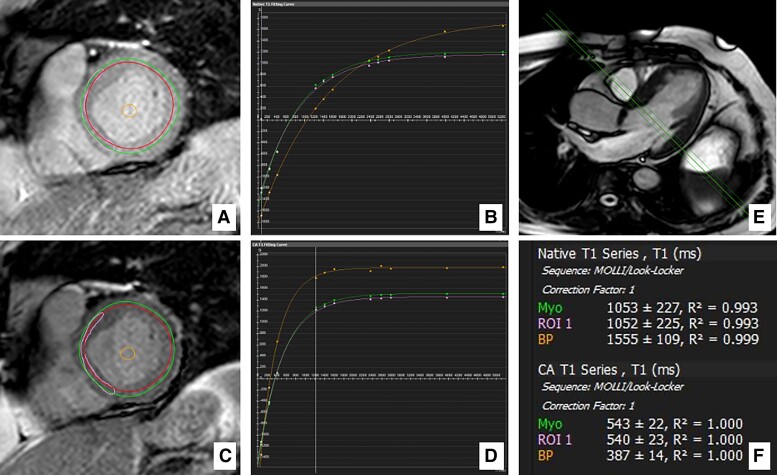
Figure 1.
Regions of interest and T1 measures. Example of basal-level short-axis region of interest selection and T1 measurements in postoperative cardiovascular magnetic resonance in a patient with severe aortic stenosis (global ECV with LGE = 26.1%; iECV = 17.4 mL/m2). (A and B) native regions of interest and T1 maps. (C and D) post-contrast regions of interest and T1 maps. (E) long-axis view showing the corresponding basal position of the (A and B) short-axis slices. (F) automatic measurement of native and post-contrast T1 times. Myo, global level selection; ROI 1, septal selection; BP, blood pool selection.

