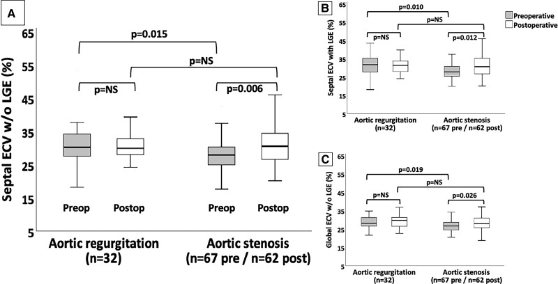
Figure 3.
Different ECV measures in patients with aortic valvular heart disease. Comparison between three different extracellular volume (ECV) measures in aortic regurgitation and stenosis on pre- and postoperative cardiovascular magnetic resonance. (A, B, and C) show the septal ECV without (w/o) late-gadolinium enhancement (LGE), septal ECV with LGE, and global ECV without LGE, respectively. Solid horizontal lines indicate median values; boxes indicate p25 and p75; and vertical lines indicate the highest and lowest values. P values indicate differences between measures (significant if < 0.05). NS, non-significant. Preop, preoperative. Postop, postoperative.

