Graphical Abstract.
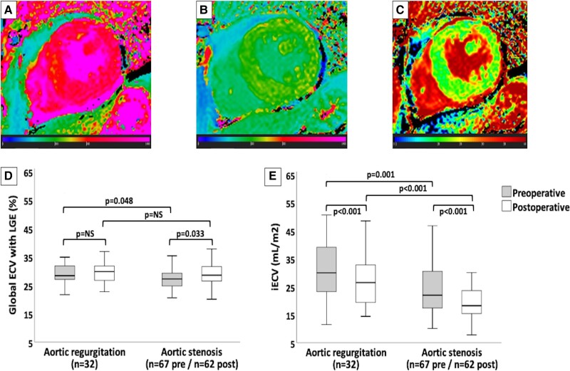
Diffuse myocardial fibrosis measures and changes in ECV and iECV. Panels A, B, and C demonstrate automatic native T1, post-contrast T1, and extracellular volume (ECV) measures, respectively, in the postoperative cardiovascular magnetic resonance of a patient with severe aortic stenosis (Global ECV with LGE = 26.1%; iECV = 17.4 mL/m2). Graphics D and E show comparisons between the pre- and postoperative measures of the global ECV with late gadolinium enhancement (LGE) and the indexed extracellular volume (iECV), respectively, in aortic valvular heart diseases. Solid horizontal lines indicate median values; boxes indicate p25 and p75; and vertical lines indicate the highest and lowest values. P values indicate differences between measures (significant if <0.05). NS, non-significant.

