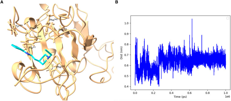
Fig. 5. Model of GEN727 in the lipid binding pocket of SARS CoV-2 RBD.
(A) Snapshot from MD simulation at the end of 1 μs. (B) Plot of protein-ligand distance [between the center of mass of GEN727 (shown in cyan/blue) and the center of mass of the lipid binding pocket, heavy atom only, in nm], as a function of simulation time (in ps). The lipid binding pocket is defined by five Phe residues, Phe338, Phe342, Phe374, Phe377, and Phe392.

