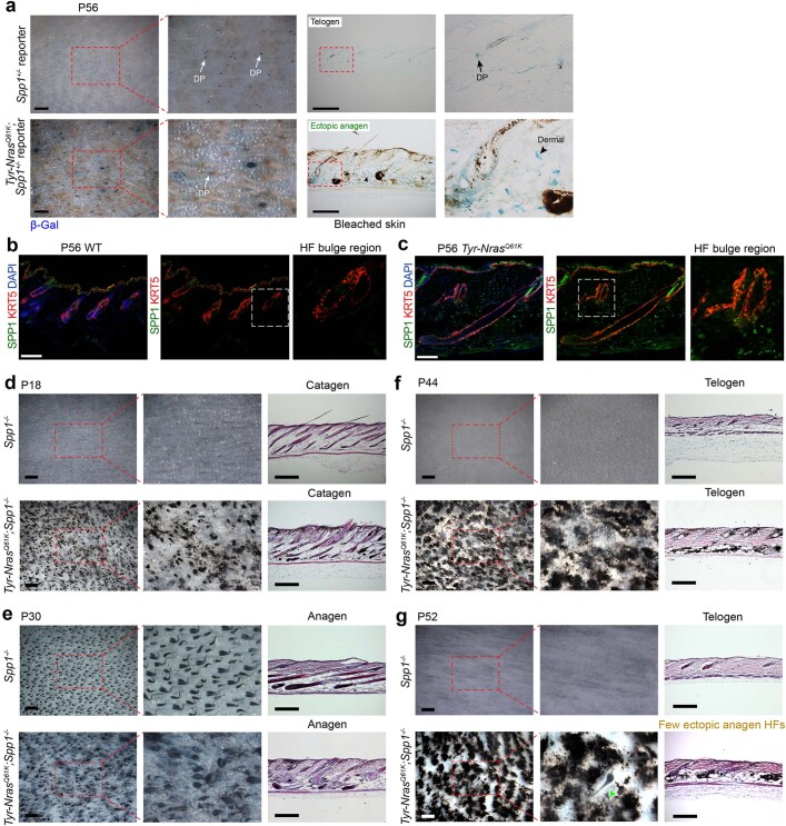
Extended Data Fig. 8. Changes in expression and the effect of osteopontin deletion in Tyr-NrasQ61K skin.
a-c, Osteopontin expression is increased in Tyr-NrasQ61K skin. a, Spp1 reporter activity was increased in Tyr-NrasQ61K skin. LacZ staining (blue) on Tyr-NrasQ61K;Spp1+/− vs. control Spp1+/− P56 reporter mouse skin showed broad increase in LacZ+ cells. Dermal and dermal papilla (DP) expression sites are marked. For each panel, representative wholemount and histology samples are shown on the left and on the right, respectively. b–c, Co-immunostaining for KRT5 (red) and SPP1 (green) in P56 WT control (b) and Tyr-NrasQ61K skin (c). Tyr-NrasQ61K skin showed prominently increased SPP1 expression in the dermal compartment, including around bulge regions of HFs (inserts). d-g, Osteopontin deletion rescues hair cycle quiescence in Tyr-NrasQ61K mice. Tyr-NrasQ61K;Spp1−/− mice showed rescue of hair cycle quiescence. Unlike Tyr-NrasQ61K mice (see Extended Data Fig. 1), Tyr-NrasQ61K;Spp1−/− mice showed synchronized catagen at P18 (d), synchronized anagen at P30 (e), synchronized telogen at P44 (f) and largely synchronized telogen P52 (g). For each time point, representative Spp1-/- control and Tyr-NrasQ61K;Spp1-/- mutant skin samples are shown. Wholemount samples are shown on the right and histology on the left of each panel. Scale bars, b, c – 100 μm; a, d–g (wholemount) – 500 μm; a, d–g (histology) – 200 μm.

