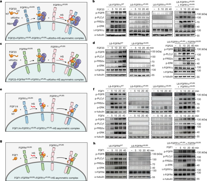
Fig. 5. Both endocrine and paracrine FGFs signal via asymmetric receptor dimers.
a, Schematic diagram showing that, in response to FGF23 and αKlotho cotreatment, FGFR1cΔSLBS and FGFR1cΔPLBS can complement each other and form a 1:1:1:1:1 FGF23–FGFR1cΔSLBS–FGFR1cΔPLB–αKlotho–HS asymmetric complex. b, Immunoblot analyses of whole extracts from untreated or FGF23- and αKlotho-cotreated L6 cell lines singly expressing FGFR1cWT, FGFR1cΔPLBS and FGFR1cΔSLBS or co-expressing FGFR1cΔSLBS with FGFR1cΔPLBS probed as in Fig. 2. c, Schematic diagram showing that FGF23, αKlotho and FGFR4ΔSLBS (serving as a primary receptor) form a ternary complex and recruit FGFR1cΔPLBS as a secondary receptor, in the presence of HS. d, FGF23- and αKlotho-cotreated L6 cell lines stably expressing FGFR4WT, FGFR4ΔSLBS alone or FGFR4ΔSLBS together with FGFR1cΔPLBS were analysed for FGFR activation/signalling using western blotting of total cell lysates as in Fig. 2. e, Schematic diagram showing that, in response to paracrine FGF1/4 and HS, FGFR1cΔSLBS and FGFR1cΔPLBS can complement each other and form 1:1:1:1 FGF–FGFR1cΔSLBS–FGFR1cΔPLBS–HS asymmetric signalling complexes. f, L6 cell lines expressing FGFR1cΔSLBS and FGFR1cΔPLBS individually or co-expressing them were treated with FGF1 (1 nM) and cell extracts were immunoblotted as in Fig. 2. g, Schematic diagram showing that FGF1, HS and FGFR4ΔSLBS (serving as primary receptor) form a stable complex which subsequently recruits FGFR1cΔPLBS as a secondary receptor. h, L6 cell lines expressing FGFR4WT or FGFR4ΔSLBS singly or co-expressing FGFR4ΔSLBS with FGFR1cΔPLBS were treated with FGF1 (1 nM) and cell extracts were immunoblotted as in Fig. 2. Experiments were performed in biological triplicates with similar results. CT, C terminus; NT, N terminus.

