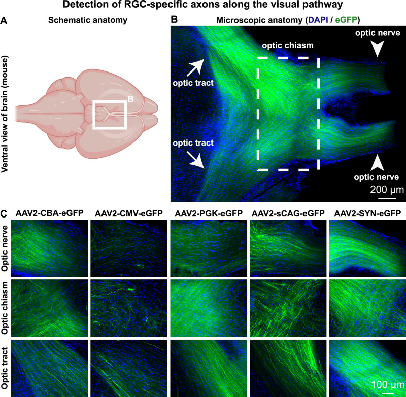
Fig. 6. eGFP-positive axons of transduced mouse retinal ganglion cells are detected at the optic nerve, optic chiasm and optic tract.
A Schematic representation of a ventral view on the mouse brain, in which the white square illustrates the location of the optic chiasm and related structures. B Microscopic overview of the optic chiasm from a mouse that had bilateral AAV2-SYN-eGFP intravitreal injections. This image is modified from the panel containing AAV2-SYN-eGFP in Fig. 3C. C eGFP-positive fibres were found at the optic nerve, optic chiasm and optic tract in all investigated experimental groups. All images were taken using a tile-scanning epifluorescence microscope with exposure settings selected to maximise axon visualisation for each promoter. Horizontal sections were stained for DAPI, eGFP is shown throughout without amplification using a GFP antibody. Schematic was created with BioRender.com.

