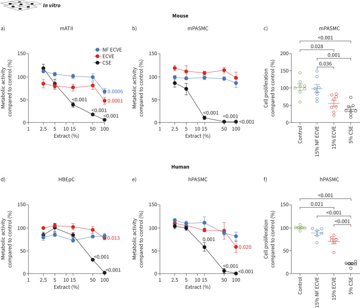
FIGURE 1.
Effect of in vitro nicotine-containing e-cigarette vapour extract (ECVE) or nicotine-free e-cigarette vapour extract (NF ECVE) exposure on metabolic activity and proliferation. a, b) Cell metabolic activity of primary mouse alveolar type II (mATII) cells (a, n=6) and primary mouse pulmonary arterial smooth muscle cells (mPASMCs) (b, n=5) after exposure to different concentrations of either NF ECVE, ECVE or conventional cigarette smoke extract (CSE). Data are presented as percentage of control. c) Cell proliferation of mPASMC (n=8) after exposure to either 15% NF ECVE, 15% ECVE, 5% CSE or control. Data are presented as percentage compared to control. d, e) Cell metabolic activity of primary human bronchial epithelial cells (HBEpCs) (d, n=6) and primary human PASMCs (hPASMCs) (e, n=5) after exposure to different concentrations of either NF ECVE, ECVE or CSE. Data are presented as percentage of control. f) Cell proliferation of hPASMCs (n=6) after exposure to either 15% NF ECVE, 15% ECVE, 5% CSE or control. Data are presented as percentage compared to control. Controls were treated with medium without ECVE, NF ECVE or CSE. Number of mATII cells, mPASMCs and hPASMCs represents independent isolations per group, number of HBEpCs represents independent experiments per group. Statistical analysis was performed by one-way ANOVA with Tukey's post hoc test. Significant p-values in comparison to respective controls are presented. Data are presented as mean±sem.

