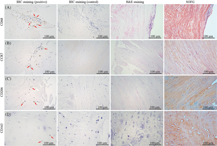
FIGURE 2.

The histological staining and immunohistochemistry of herniated nucleus pulposus (NP) derived from patients with lumbar disc hernia (LDH). The hematoxylin and eosin (H&E) and Safranin O/Fast Green (SOFG) staining showed that NP tissues developed structural abnormalities, such as extracellular matrix disorder, with cracks and tears, reduced cell counts, and cell cluster formation. The accumulation of macrophage markers CD68, CCR7, CD206, and CD163 was detected in NP samples of LDH patients (red arrow).
