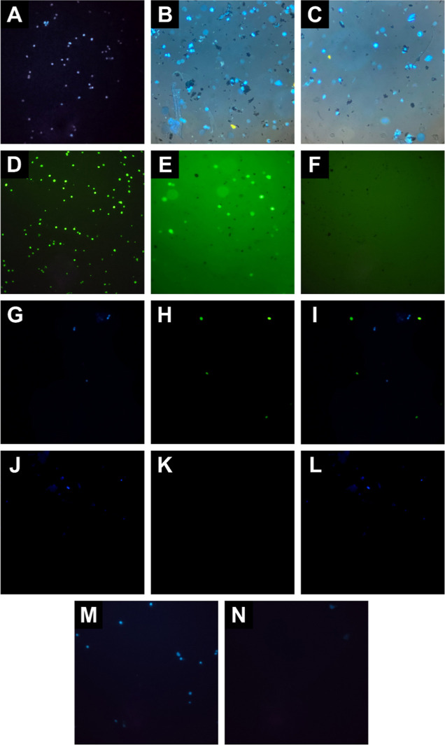
Figure 2.
Representative fluorescent microscopy images of off-chip cell isolation, (A) SKBR3 cells stained with DAPI, (B,C) stained SKBR3 cells incubated with HER2-MNPs before and after magnetic washing, (D) MDA-MB-231 cells stained with fluorescein dye, (E,F) stained MDA-MB-231 cells incubated with HER2-MNPs before and after magnetic washing, (G–I) blue, green, and merged fluorescent images of stained SKBR3 and MDA-MB-231 cell cocktail incubated with HER2-MNPs before magnetic washing, (J–L) blue, green, and merged fluorescent images of stained SKBR3 and MDA-MB-231 cell cocktail incubated with HER2-MNPs after magnetic washing, aand (M,N) stained SKBR3 cells incubated with bare MNPs before and after magnetic washing.

