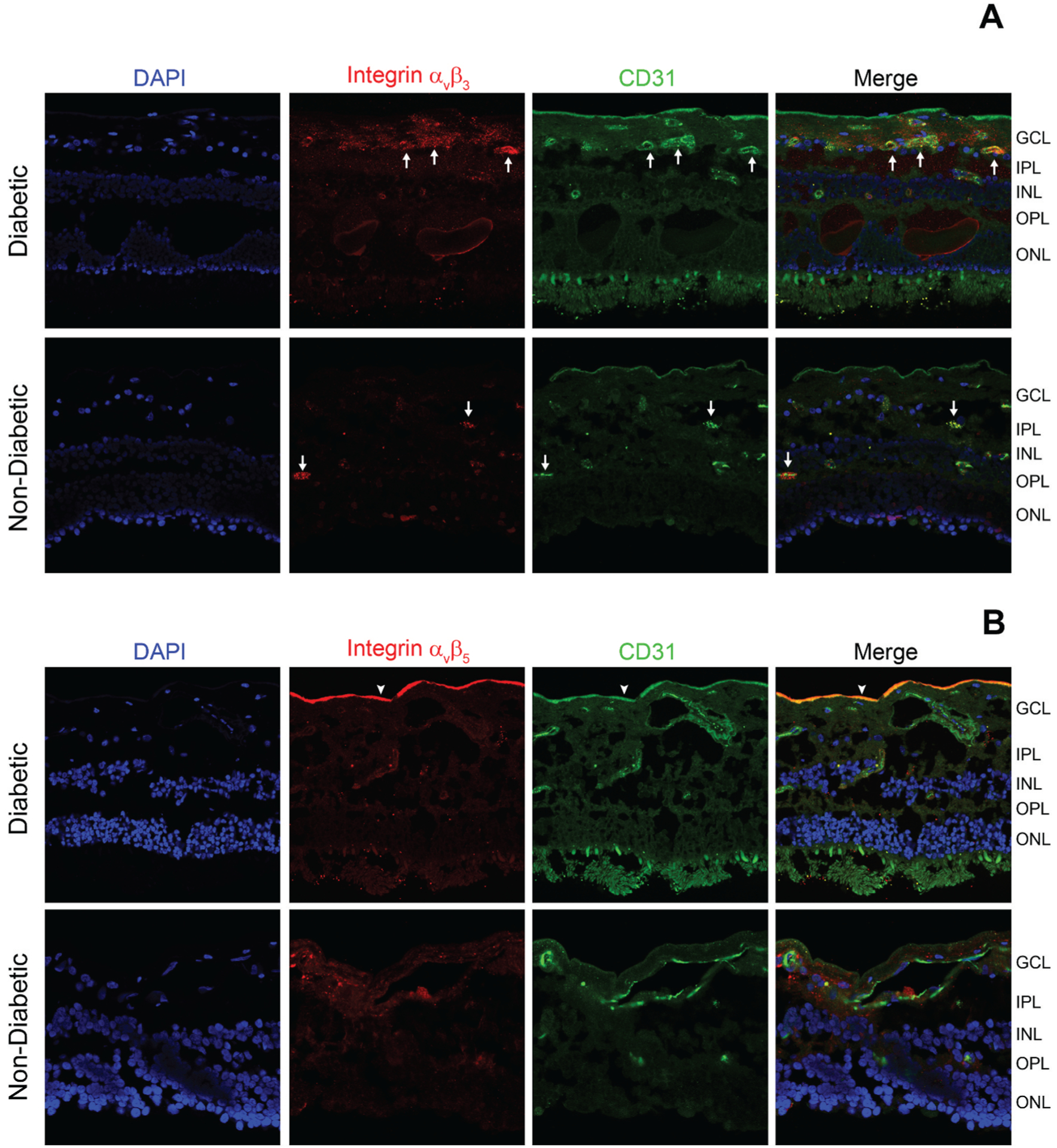
Figure 1.

Frozen ocular sections of human donors (non-diabetic and diabetes with macular edema with non-proliferative diabetic retinopathy (NPDR)) were stained with DAPI (highlighting cell bodies), integrin αvβ3 or αvβ5 (red, Millipore MAB1976 or MAB1961, 1:100) antibodies, and CD31 antibodies (highlighting endothelial cells, green, Abcam ab28364, 1:100). (A) Representative photomicrographs are showing an increase in the staining for αvβ3 in the ganglion cell layer and around the vessels (white arrows) from a diabetic donor (B) Representative photomicrographs are showing more intense αvβ5 staining near the internal limiting membrane (arrowhead) in a diabetic donor. GLC-ganglion cell layer, IPL-inner plexiform layer, INL-inner nuclear layer, OPL-outer plexiform layer, ONL-outer nuclear layer.
