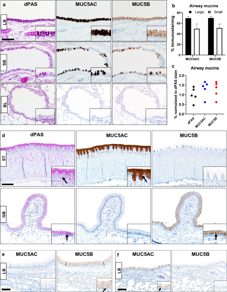
Fig. 1.
Mucin detection in ferret tissues. a Representative mucin detection (insets) by dPAS, MUC5AC and MUC5B in surface epithelia of large bronchi (LB) or small bronchi (SB) and bronchioles (BL) from WT ferrets (bar = 130 µm). b Evaluation of MUC5AC and MUC5B immunostaining in surface epithelia of large and small bronchi (N = 5 WT ferrets) using digital image analysis. Airway size was a significant factor for the variance in mucin detection (P = 0.0072, Two-way ANOVA). c Comparison of dPAS, MUC5AC and MUC5B in serial sections of WT bronchus (N = 5 WT ferrets) showed no significant differences in mucin detection between stain method (P = 0.2938) Kruskal–Wallis test). d Representative images of differential mucin expression (arrows and insets) in WT ferret stomach (ST, bar = 170 µm) and gallbladder (GB, bar = 85 µm). dPAS, MUC5AC and MUC5B. e Mucin detection (arrow and insets) in a large bronchus (LB) from a MUC5AC−/− ferret (N = 1). Surface epithelia and submucosal glands were MUC5B + confirming the tissue was viable for immunostaining. MUC5AC immunostaining was absent from the surface epithelia, and this was consistent with the ferret’s genotype, bar = 85 µm. f Mucin detection (inset and arrows) in a large bronchus (LB) from a MUC5B−/− ferret (N = 1). The surface epithelia of the bronchus was MUC5AC+ confirming the tissue was viable for immunostaining, but it was MUC5B- consistent with the tissue genotype, bar = 85 µm

