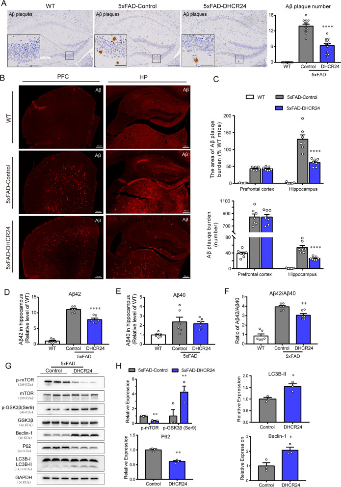
Fig. 4.
DHCR24 knock-in alleviates Aβ pathology and activates autophagy flux in hippocampus of 5xFAD mice. A The immunohistochemistry images of Aβ plagues using the antibody MOAB-2 in the hippocampus (Scale bars, 200 μm). Insets show a higher magnification of Aβ plagues. Inset scale bar, 50 μm. The right is analysis of number of Aβ plagues in hippocampus. n = 12 slices from 4 mice per group. B Fluorescence images of Aβ (red) in prefrontal cortex and whole hippocampus (scale bar, 200 μm). C The top half is the analysis of the area of Aβ plagues, and the bottom half is the analysis of number of Aβ plagues in prefrontal cortex and hippocampus. n = 7 slices from 4 mice per group. D, E The relative level of Aβ42 and Aβ40 in hippocampus by ELISA which normalized with WT mice. n = 6 mice per group. F The ratio of Aβ42/Aβ40 in hippocampus which normalized with WT mice. G The immunoblotting bands of p-mTOR, p-GSK3β (ser9), Beclin-1, P62 and LC3B in the hippocampus of 5xFAD-Control group and 5xFAD-DHCR24 group. H Analysis of western blot with mean gray value which all were quantification on the ratio of target proteins against GAPDH except p-mTOR and p-GSK3β (ser9) was against total mTOR and GSK3β, n = 3 mice per group. Data expressed as mean ± SEM, statistical analysis between two groups was analyzed by unpaired two-tailed Student’s t-test, between three groups was analyzed by one-way ANOVA with Tukey’s post hoc test. *P < 0.05; **P < 0.01; ***P < 0.001; compared with aged-matched 5xFAD-Control group

