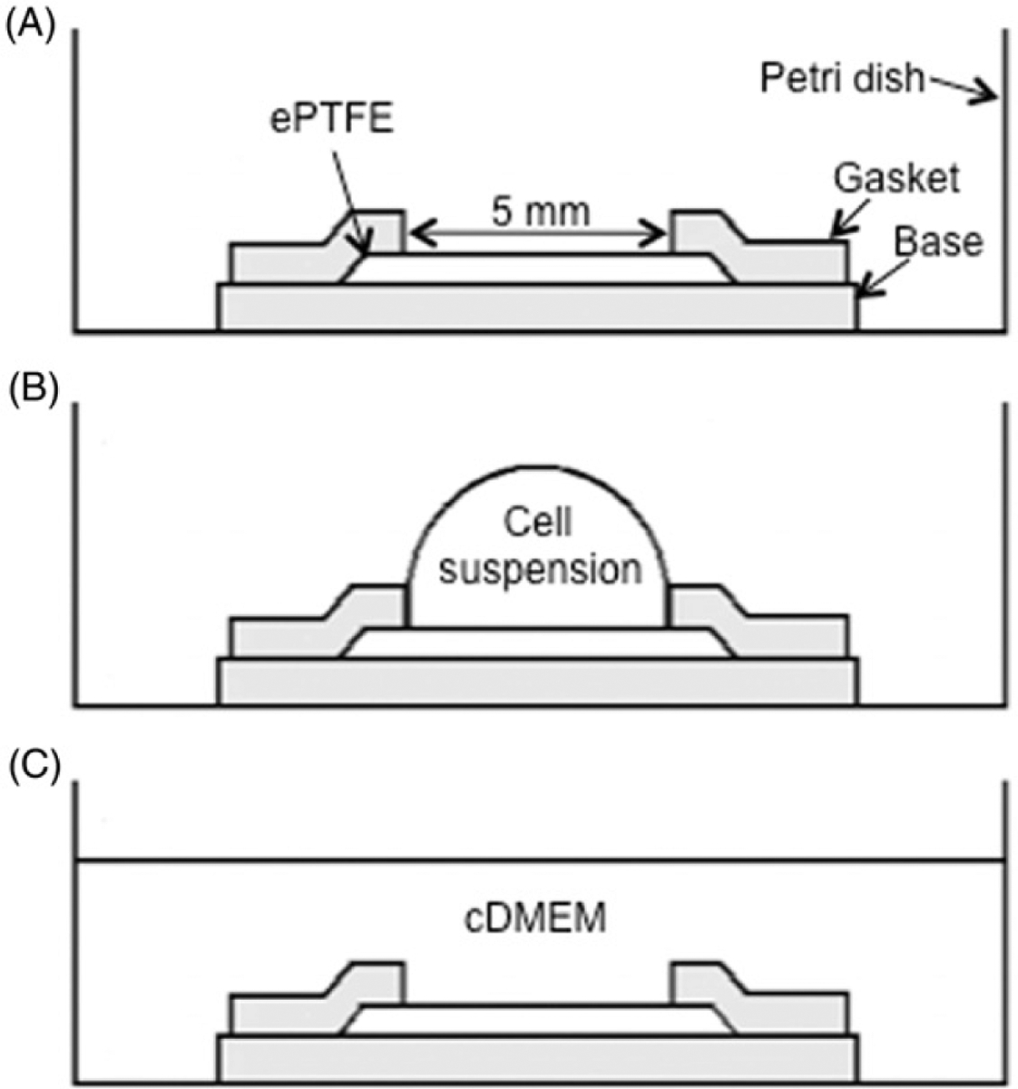
Figure 1.

Sketch of the cell seeding process (side view). (A) The 5 mm×5 mm ePTFE sheet was held in place by a silicone holder’s base and gasket (shaded in the diagram) in a standard Petri dish for tissue culture. (B) Cell suspension was pipetted onto the ePTFE sheet. (C) 2.5 mL of cDMEM was added to the dish. Drawings not to scale.
