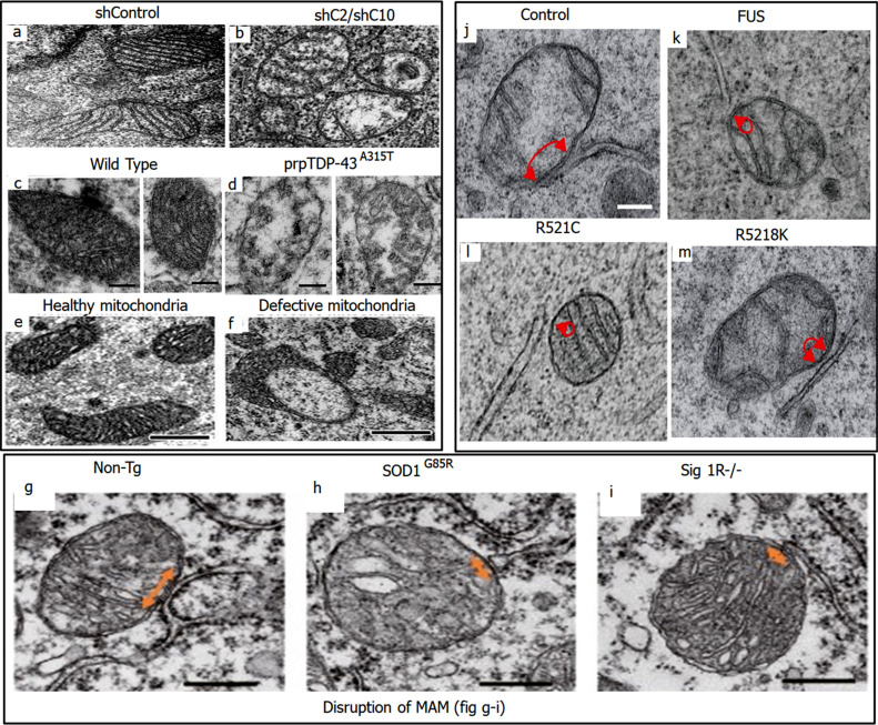
Fig. (3).
Morphological changes observed in mitochondria. (a and b) depict normal and abnormal cristae in mitochondria of control and CHCHD1/CHCHD10 silenced HeLa cells respectively. Reproduced from ‘Cell Death and Disease’, Volume 13, authored by Yu Ruan, et al. CHCHD2 and CHCHD10 regulate mitochondrial dynamics and integrated stress response. Copyright © The Author(s) 2022. Published by Nature; License Link: (https://creativecommons.org/licenses/by/4.0/). (c and d) depict the defects in mitochondria in TDP43 pathology within the corticospinal motor neurons (CSMN) of WT and prpTDP-43A315T mice, respectively. Reproduced from ‘Scientific Reports’, Volume 12, authored by Mukesh Gautam, et al. [92] Mitochondrial dysregulation occurs early in ALS motor cortex with TDP-43 pathology and suggests maintaining NAD+ balance as a therapeutic strategy. Copyright © 2022, The Author(s). Published by Nature; License Link: (https://creativecommons.org/licenses/by/4.0/). (e and f) depict the healthy and defected mitochondria respectively in Corticospinal motor neurons (CSMN) at P15. Reproduced from ‘Cellular Neuropathy’, Volume 13, authored by Mukesh Gautam, et al. [93] Mitoautophagy: A Unique Self-Destructive Path Mitochondria of Upper Motor Neurons With TDP-43 Pathology Take, Very Early in ALS. Copyright © 2019 Gautam, Xie, Kocak and Ozdinler. Published by Frontiers in Cellular Neuroscience; License Link: (https://creativecommons.org/licenses/by/4.0/). (g-i) depict the MAM in WT, SOD1G85R and SigR1 knockout mice model respectively. The arrows represent MAM area, and it is clearly observed that MAM area is reduced in SOD1G85R and SigR1. Reproduced from ‘EMBO Molecular Medicine’, Volume 8, authored by Seiji Watanabe, et al. [90] Mitochondria-associated membrane collapse is a common pathomechanism inSIGMAR1- andSOD1-linked ALS. Copyright © 2016 The Authors. Published by EMBO Press; License Link: (https://creativecommons.org/licenses/by/4.0/). (j-m) also depict the MAM area in NSC34 cells transfected with CTRL, WT and FUS mutated gene vectors. The arrows represent the MAM area. Significantly reduced MAM area is observed in WT as well as mutant FUS expressed cells. Reproduced from ‘EMBO Reports’, Volume 17, authored by Radu Stoica, et al. [51] ALS/FTD-associated FUS activates GSK‐3β to disrupt the VAPB–PTPIP51 interaction and ER–mitochondria associations. Copyright © 2016 The Authors. Published by EMBO Press; License Link: (https://creativecommons.org/licenses/by/4.0/).

