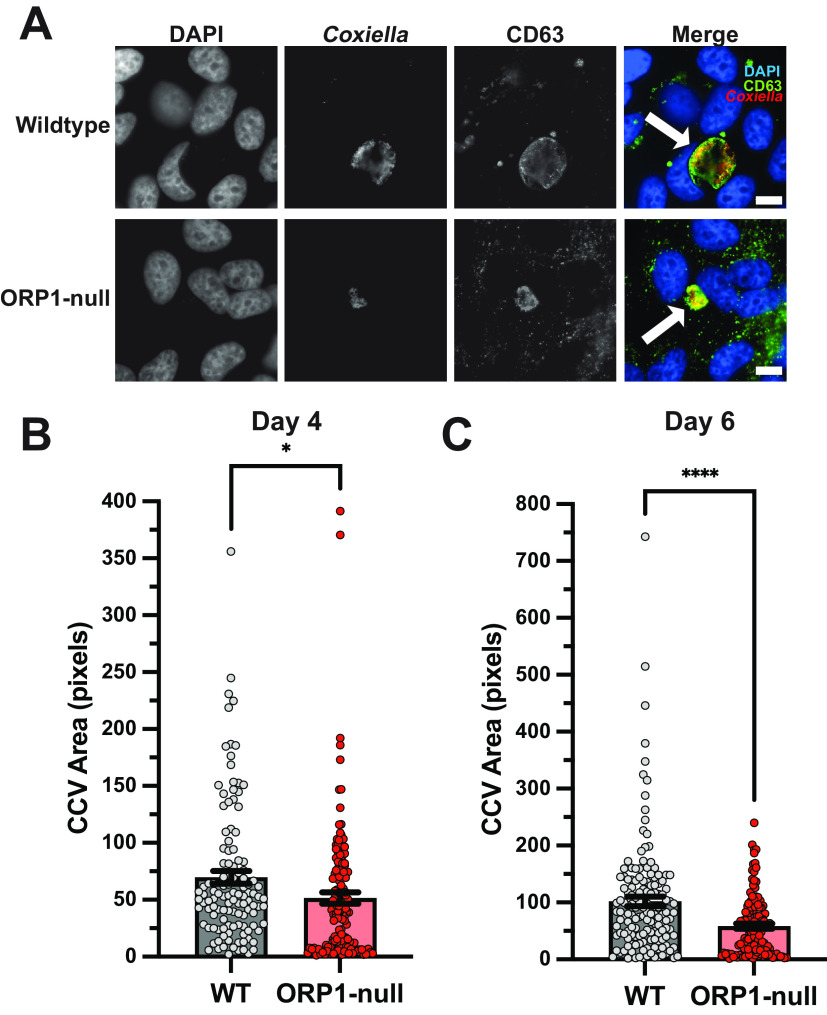
FIG 2.
CCV expansion is attenuated in ORP1-null HeLa cells. (A) Representative images of wild-type (WT) and ORP1-null HeLa cells infected with mCherry-expressing C. burnetii for 6 days, then immunofluorescence stained with anti-CD63 antibodies and DAPI. White arrows indicate CCVs. Scale bars, 10 μm. (B) CCV areas in WT and ORP1-null cells after 4 days of infection with mCherry-expressing C. burnetii. WT CCV mean area was 69.65 pixels, and ORP1-null CCV mean area was 51.55 pixels. (C) CCV areas in WT and ORP1-null HeLa cells after 6 days of infection with mCherry-expressing C. burnetii. CCV mean area in WT cells was 101.9 pixels, and ORP1-null CCV mean area was 58.34 pixels. Data shown are means ± standard errors of the mean (SEM) of at least 20 cells per condition in each of three independent experiments as determined by unpaired Student's t test. *, P < 0.05; ****, P < 0.0001.

